کچھوؤں کے لیے ایکسرے۔ کیسے، کہاں، کیسے سمجھیں؟

X-rays can be done in any clinic or veterinary clinic equipped with an X-ray machine.
Why is an x-ray done? 1. Check for pneumonia (pneumonia) 2. Check for foreign bodies in the stomach of turtles or eggs in females. 3. See if there is a fracture of the limb.
Average shooting parameters (for small and medium):
If the picture is an overview, then from a distance of about 90 cm, the shooting parameters are approximately 40-45 kV and 6-12 mas.
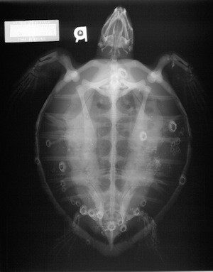
For an adult ruby to see the eggs: about 50 kV at 10 mA. If there is a suspicion that the egg shell is poorly formed, then the shooting mode is 45-50-55 kV / 10-15mAs. Eggs and intestinal patency are viewed in a dorso-ventral projection.
When diagnosing fractures: 40-45 kV and 6-12 mA
The larger the turtle, the “harder” the shot. For a medium-sized Central Asian woman, the “average” mode is 40kV x 6-10 mAs.
For small water and land animals with suspicion of an x-ray detectable foreign body or obstruction: two x-rays, in dorso-ventral (from the back) and lateral projection, shooting mode approximately 40kV x 10-15 mAs (this is for the radiologist). Ideally, if 45 minutes before the shooting, 10% barium sulfate is injected into her stomach, somewhere around 5-7 ml, diluted with starch broth (this is with obstruction). For radiopaque images, use omnipaque, barium sulfate, or at least urographin (as for urography). Urografin 60% is diluted with water twice and 15 ml / kg of solution is injected. The contrast is injected into the stomach with a probe. If obstruction is suspected, two pictures are taken – one hour later and 6-8 hours or 24 hours later – or one 24 hours after the contrast injection. The most important image is dorso-ventral. The side is not required and most often not needed, there you already need to look at the situation.
Suspicion of pneumonia: In the usual projection (dorso-ventral), internal organs are projected onto the lung fields, and instead of the lungs, only their fragments are visible. Pneumonia in turtles is established only in the cranio-caudal projection, and in the lateral one – an auxiliary image. It makes sense only for large and medium-sized turtles, at least from 12 cm. For small ones, it will be uninformative.
If you need to see what’s wrong with the jaw joint: An x-ray is needed, but with a very good resolution (for example, on a mammograph). It is best to make the animal lightly anesthetized and try to open its mouth under anesthesia. If this fails, insert something like a bar as a mouth expander and take a picture in the lateral and dorso-ventral projection with the jaws as open as possible.
Some of the pictures are taken from spbvet.com
کچھی صحت کے دیگر مضامین
© 2005 - 2022 Turtles.ru


آپ کو بھی پسند فرمائے
درخت، جھاڑیاں - کچھوؤں کے بارے میں اور کچھوؤں کے لیے
Helminthiases: گول کیڑے، آکسیورائڈز اور دیگر کیڑے